Radiology and Imaging
The goal of the Tift Regional Medical Center Radiology Department is to offer advanced medical imaging while assuring the comfort of all patients. The results of our diagnostic exams are interpreted by local, experienced radiologists who work with physicians to reach the diagnosis needed to best treat patients. Urgent cases can be scheduled the same day. For more information on Radiology services at Tift Regional Medical Center, please call 229-353-7500 or toll free 800-648-1935, ext. 7500.
RadiologyInfo, a website sponsored by the American College of Radiology and the Radiological Society of North America, has more information related to the many radiologic procedures and therapies available to you and your family. RadiologyInfo tells you how various x-ray, CT, MRI, ultrasound, radiation therapy and other procedures are performed. It also addresses what you may experience and how to prepare for the exams. Please click here.
Radiology and Imaging Services
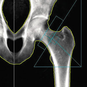
To accurately detect osteoporosis, doctors commonly use DEXA bone densitometry scan to safely and painlessly measure bone density and bone loss by taking a low dose x-ray of your lower spine and hips.
Sometimes, an additional low-dose x-ray image, called VFA (Vertebral Fracture Assessment), is performed of the entire spine. This test allows doctors to see existing vertebral fractures, which may indicate the need for more aggressive treatment, even if bone density results are in the “normal” range.
When doctors detect bone loss in the earliest stage, treatment, along with preventative measures, are more successful.
The DEXA test also assesses your risk for developing fractures. It is effective in tracking the effects of treatment for osteoporosis and other conditions that can cause bone loss. Bone density testing is recommended for:
- Post-menopausal women age 60 or older who have risk factors for developing osteoporosis
- Patients with a personal or maternal history of hip fracture or smoking
- Post-menopausal women who are tall (over 5 feet 7 inches) or thin (less than 125 pounds)
- Men and women who have hyperparathyroidism
- Men and women who over an extended period have taken medications that are known to cause bone loss
How should I prepare for this procedure?
Before:
- Refrain from taking calcium supplements for at least 24 hours beforehand
- Wear comfortable clothing and avoid garments that have zippers, belts or buttons made of metal
- Inform your technologist if you’ve recently had a barium examination or have been injected with a contrast material for a CT or radio-isotope scan
- Inform your technologist if there is a possibility you may be pregnant
What happens during the bone densitometry exam?
During:
DEXA bone densitometry is a simple, painless, and non-invasive procedure. During your exam, you will lie on a padded table while the bone densitometry system scans two or more areas, usually your hip and spine. Unlike typical x-ray machines, radiation exposure during bone densitometry is extremely low. The entire process takes only minutes to complete. It involves no injections or invasive procedures, and if you have no zippers or metal buttons on your clothing, you can remain fully clothed. However, all women are required to remove only their bras prior to taking the exam.
Depending on the equipment used and the parts of the body being examined, the test takes approximately 20 minutes. You’ll lie on a padded table with an x-ray generator below and a detector (an imaging device) above. It is important that you remain as still as possible during the procedure to ensure a clear and useful image.
Who interprets the results and how do I get them?
After:
The results of a DEXA bone density exam are interpreted by a radiologist and forwarded to your doctor. However, some doctors may prefer to interpret their patients’ scans. Your test results will be in the form of two scores:
T score – This number shows the amount of bone you have compared to a young adult of the same gender with peak bone mass. A score above -1 is considered normal. A score between -1 and -2.5 is classified as osteopenia: the first stage of bone loss. A score below -2.5 is defined as osteoporosis. It is used to estimate your risk of developing a fracture.
Z score – This number reflects the amount of bone you have compared to other people in your age group and of the same size and gender. If it is unusually high or low, it may indicate a need for further medical tests.
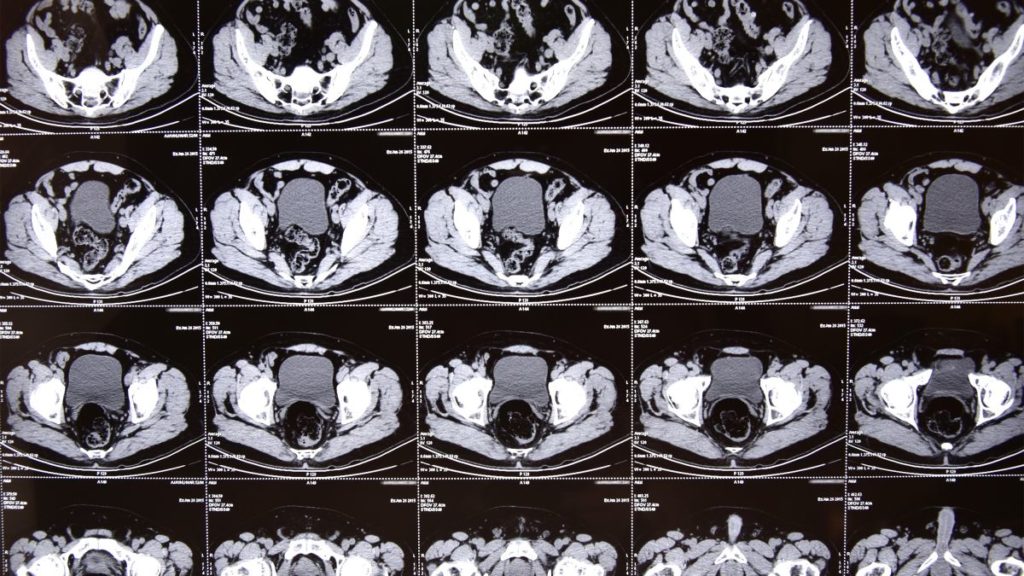
Tift Regional offers two state-of-the-art scanner suites that provide the latest in ultra-modern CT scanning. CT, or computerized tomography, is a doughnut-shaped scanning device that takes images of cross-sections of the body. CT can see into areas that cannot be seen on regular X-ray examinations. With Tift Regional’s advanced GE Lightspeed Scanner, images are made six times faster than traditional CT scanners. Reducing the scan time significantly can allow physicians to begin treating the patient more quickly.
Smart Score
The prevention of heart attacks takes another step forward at Tift Regional Medical Center. Smart Score is designed to check for risks of cardiovascular disease (CVD) and heart attacks in people who haven’t had any heart problems in the past. This test is able to help many people before they ever experience any problems associated with cardiovascular disease and heart attacks. The test shows calcification of the arteries with a simple CT scan.
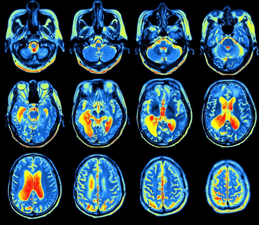
Positron Emission Tomography, also called a PET scan, is a Nuclear Medicine exam that produces a 3D image of functional processes in the body.
Positron Emission Tomography (PET) scans use radio-active tracers injected intravenously (IV) to obtain images of the human body’s function and reveal information of health and disease. As the tracers journey through the body, the scanner records signals that the tracers emit and the targeted organs collect. A computer then interprets the signals and merges them with CT images, which reveal biological maps of organ function.
This type of imaging is some of the most advanced available to diagnose cancers and other diseases. PET scans are also utilized to examine the effects of cancer therapy by characterizing biochemical changes in the cancer. To determine if they are candidates for surgery, patients who have suspected or proven brain tumors, or who have seizure disorders that are not responsive to medical therapy can be evaluated using brain scans. Patients who have memory disorders of an undetermined cause are not candidates for surgery.
Patients lie on a table that moves slowly through the large opening of the PET scanner. Scan times usually average about 30 minutes, but vary depending on the region being assessed.
Before:
You should:
- Wear comfortable clothes with no metal zippers or buttons
- Avoid exercise and not engage in any form of strenuous or vigorous activity for 24 hours before your exam; for example, golfing, swimming, heavy lifting and housework
Any additional preparation for this test varies depending on the purpose of the scan. Expect a call from our preregistration staff who will instruct you on how to prepare for your exam.
During:
- Plan on being at the office for 1 ½ to 2 hours (registration, injection and scan duration are included in the time approximation)
- You will receive an intravenous (IV) injection of the radio-active pharmaceutical
- The radio-active tracer will take approximately 60 minutes to travel through your body and be absorbed by the tissue under study. During this time, you will be in a comfortable recliner and private room. You will be asked to rest quietly and avoid significant movement or talking, which may alter the localization of the administered substance
- You will be positioned on the PET scanner table and asked to lie still during your exam
- The table will move slowly through the large opening of the PET scanner
- Scanning of the brain takes less than ten minutes. Scanning of the body takes between 15 and 25 minutes
- Usually, there are no restrictions on daily routine after the test. You should drink plenty of liquids to flush the radio-active substance from your body
What will I experience during the procedure?
- When given the intravenous injection, you will feel a slight prick. However, you will not feel the substance in your body
- You will be made as comfortable as possible on the exam table before you are positioned in the PET scanner for the test
- Your technologist can see, hear and speak with you at all times during your scan
After:
The small amount of radiotracer in your body will lose its radio-activity over time through the natural process of radio-active decay. Following the test, it may also pass out of your body through your urine or stool during the first few hours or days. You should not feel any side effects and be able to return to your normal daily activities directly after the scan. We recommend that you hydrate thoroughly for 48 hours after the scan to help void the medications.
Keep your distance from children and pregnant women for approximately six hours after a PET scan.
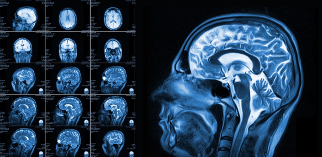
Southwell facilities offers both full-sized, high-field magnetic resonance imaging (MRI) and open MRI. Each utilize the physical properties of magnetic fields, radio waves, and computers to generate images of the body. MRI imaging is used for virtually all parts of the body. For claustrophobic patients, small children or larger patients who may find the traditional MRI too confining, the open MRI unit is available, providing a “wall-less” environment.
Magnetic Resonance Imaging (MRI) is a non-invasive test using detailed images to diagnose medical conditions. MRI uses a powerful magnetic field, radio waves and a computer to produce detailed pictures of internal body structures. MRI does not use radiation (x-rays).
Before:
MRI uses strong magnets, so it is important that you notify your doctor of any metal that may be implanted into your body. Jewelry should be left at home. If required, you’ll be asked to remove your watch, hearing aid, and other metal objects. Some makeup also contains traces of metal, so you might have to remove that, too. Braces and fillings normally aren’t a problem. You will be asked to change into a gown. Many clothing items contain metals that could potentially heat up and cause burns. Gowns are provided as well as secured lockers for valuables.
Notify your technologist if you have:
- Any prosthetic joints, such as hip and/or knee
- A heart pacemaker (or an artificial heart valve), a defibrillator or an artificial heart valve
- An intrauterine device (IUD)
- Any metal plates, pins, screws, or surgical staples in your body
- Any previous brain surgery
- Tattoos and permanent make-up
- A bullet or shrapnel in your body, had metal removed from your eye or ever worked around grinding metal
- Any possibility that you may be pregnant or suspect you may be pregnant
- Claustrophobia and require a sedative. Please ask your referring physician to prescribe one for you
In many cases, patients with a pacemaker cannot have an MRI (your technologist can verify if you have a ‘safe’ pacemaker). Metal used in orthopedic surgery pose no risk during an MRI (in most cases). You will also be asked if you have ever worked with metal. If there is a possibility of metal shrapnel in the eyes, you will be asked to do an x-ray prior to the MRI.
Some scans require the patient to receive an injection of gadolinium: a contrast medium. If this is the case, it will be discussed with you before the procedure. This contrast medium has a lower risk of allergic reaction or kidney damage compared to other mediums commonly used for CT scans. The amount of the contrast injected is determined by the patient’s weight.
- Preparation for MRI Pancreas or MRCP: Nothing by mouth for three hours prior to exam time
- Preparation for MRI Pelvis: Drink plenty of fluids to ensure that you have a full bladder
- Preparation for enterography: Nothing by mouth after midnight. You must arrive 45 minutes early to drink an oral contrast
What should I expect?
During:
MRI is painless. Some patients may experience a “closed in” feeling, although in most cases, our wide bore MRI scanners have alleviated this reaction. Plan on being with us for a minimum of 30 minutes, depending on the part of the body being scanned. You will be asked to remain still during the actual imaging process. You will hear loud tapping or thumping during the exam. Earplugs or earphones and your choice of music will be provided, if you choose.
Depending on the part of the body being examined, a contrast material called gadolinium may be used to enhance the visibility of certain tissues or blood vessels. A small needle is placed in your arm or hand vein and a saline solution IV drip will run through the intravenous line to prevent clotting. About two-thirds of the way into your exam, the contrast material is injected.
After:
You may return to normal activities as soon as the scan is complete. The radiologist will determine if there are any areas of concern in the internal organs or bone structures. The radiologist’s interpretation will then be available to your referring physician 48 hours after the exam. In most cases, your referring physician will discuss the results with you.
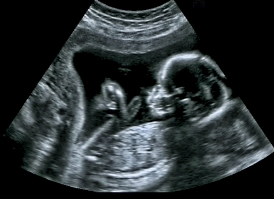
Ultrasound imaging, also called sonography, is a method of obtaining diagnostic images from inside the human body through the use of high-frequency sound waves. Ultrasonography is used as a diagnostic tool that can assist doctors with making recommendations for further treatment.
Southwell locations offer the latest ultrasound technologies that utilize high-frequency sound waves to produce moving images of the organs and structures of the body. This new generation of ultrasound equipment provides images of very high resolution and diagnostic accuracy. It is routinely used to evaluate fetal growth and complications of pregnancy. Vascular ultrasound provides accurate assessment of arteries and veins in the extremities and neck.
Pelvic Ultrasound
Pelvic ultrasound uses sound waves to form pictures of your organs that appear on a screen. It can help your physician assess pain or other symptoms in the pelvis (lower abdomen). For pregnant women, ultrasound is used to check the fetus. The test is done by moving a probe over the abdomen and pelvis. At times, it is also done by placing a probe inside the vagina. This test involves no radiation and is harmless.
How should I prepare for a Pelvic Ultrasound?
Wear comfortable, loose-fitting clothing. For a pregnancy or pelvic ultrasound, you are required to have a full bladder. To ensure best results, please follow these instructions:
- Two hours before exam time, empty your bladder.
- Do not urinate again until your exam is complete.
- Immediately after your last urination, begin drinking 1.5 quarts of liquid (no milk or dairy products). Drink at a rate that is comfortable for you.
- Please remember not to urinate until after your ultrasound exam is completed.
What should I expect during this procedure?
The examination usually takes less than 30 minutes. After being positioned on the exam table, a clear gel is applied in the area being examined. This helps the transducer make contact with the skin. The technologist firmly presses the transducer against the skin and moves it back and forth to image the area of interest.
Generally, the technologist is able to review the ultrasound images in real-time or, when the examination is complete and the gel is wiped off, you may be asked to dress and wait while the ultrasound images are reviewed, either on film or monitor.
Breast Ultrasound
Ultrasound imaging of the breast uses sound waves to produce pictures of the internal structures of the breast.
It is primarily used to help diagnose breast lumps or other abnormalities your doctor may have found during a physical exam, mammogram or breast MRI. Ultrasound is safe, non-invasive and does not use radiation.
Before:
This procedure requires little to no special preparation. Leave jewelry at home and wear loose-fitting, comfortable clothing. You will be asked to undress from the waist up and to wear a gown during the procedure.
During:
After being positioned on the exam table, a clear gel is applied in the area being examined; it may feel a bit cold. The gel helps the transducer make contact with the skin. The technologist firmly presses the transducer against the breast and moves it back and forth to image the area of interest.
After:
After a radiologist analyzes the images, preliminary results will be given to you during your visit with us. A full report will then be sent to your referring physician.
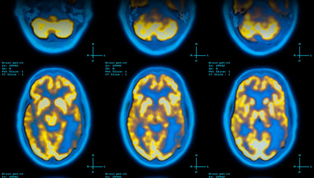
Nuclear Medicine scans use a small amount of a radio-active substance to produce 2- or 3D images of body anatomy and function. Diagnostic images produced by a nuclear scan are used to evaluate a variety of diseases. Sometimes a nuclear scan is combined with a CT scan.
What are some common uses of Nuclear Medicine?
Nuclear medicine images can assist the physician in viewing, monitoring or diagnosing:
- Tumors
- Blood flow and function of the heart
- Respiratory and blood-flow problems in the lungs
- Organ function of the kidney, bowel, gallbladder and others
Before:
Preparation for your exam varies depending on which part of your body is to be scanned. If the exam is to evaluate the stomach, you may be required to refrain from eating before the test. If the exam is to evaluate the kidneys, you may need to drink plenty of water before the test. The individual who schedules your exam will explain the preparation process.
The day before your appointment, a staff member from our office will call you to confirm. If you need to cancel or reschedule, it must be done no later than one day before your appointment. The radio-active substance used in your test is a special-order and cannot be canceled within less than 24 hours prior to your exam.
Generally, you are not required to change out of your clothes for a Nuclear Medicine procedure. However, for some studies, you are required to remove metal objects, such as loose change, pocket knives, belt buckles and some jewelry. As with any study in radiology, you should tell the technologist if there is a chance that you are pregnant or if you are breastfeeding.
During:
Although imaging time can vary, the exam generally takes between 20 and 60 minutes.
- A radiopharmaceutical, known as a tracer, either is administered intravenously or orally. The type of radiopharmaceutical used and if the imaging is conducted immediately or several hours later depends upon the type of exam you’re undergoing
- For most nuclear scans, you lie on a table and a nuclear imaging camera captures the image of the examined area. The camera either is suspended over or below the exam table or in a large donut-shaped machine similar to a CT scanner. While the images are being captured, you must remain as still as possible
- You may hear low-level or buzzing noises from the machine
- Most of the radio-activity is expelled from your body in urine or stool. The rest simply disappears over time
After:
You should not feel any side effects and be able to return to your normal daily activities directly after the scan. We recommend that you hydrate thoroughly for 48 hours after the scan to help void the medications.
VenaCure EVLT: non-surgical laser treatment available for varicose veins
Radiologist Carl Stalvey, M.D. is now offering a non-surgical laser procedure at Tift Regional Medical Center (TRMC) to treat varicose veins. If you are one of more than 25 million Americans who suffer from varicose veins, you may benefit from this treatment that’s safe, virtually painless and requires little-to-no downtime.
Varicose veins can be unsightly, uncomfortable and a symptom of vein disease. The VenaCure EVLT laser vein treatment procedure utilizes laser energy to achieve proven results without the discomfort and lengthy recovery experienced through surgical options. The entire procedure takes about an hour at Tift Regional Medical Center, and depending on the severity of the condition, may be covered by insurance.
Is VenaCure EVLT right for me?
The Venacure EVLT procedure uses a minimally invasive endovenous laser to treat varicose veins and is a clinically proven alternative to traditional and painful ligation and stripping surgery. It requires no general anesthesia and offers minimal risk and shorter recovery time.
The VenaCure EVLT procedure uses the latest generation of EVLT® catheter, which has been cleared by the Food and Drug Administration (FDA) for use in the United States, to insert a laser fiber directly inside the faulty vein under local anesthesia. The laser delivers a precise dose of energy into the vein wall, collapsing it. This process, called ablation, cures the condition and diverts blood flow to nearby functional veins. The resulting increased circulation significantly reduces the symptoms of varicose veins and improves their surface appearance.
The VenaCure EVLT treatment generally takes less than one hour in a doctor’s office and offers immediate relief with minimal-to-no scarring. Patients can usually resume normal activities immediately with minimal or no pain. The success rate of the VenaCure EVLT procedure is as high as 98 percent – measurably better than the alternatives of surgical ligation or stripping and radiofrequency electrosurgery.
When used to treat medical symptoms such as swelling and poor circulation, VenaCure EVLT treatment is usually reimbursed by health insurance. Contact your insurer to determine coverage.
Talk to your provider today. A referral from a physician, nurse practitioner or physician assistant is required. For more information, please call the TRMC Radiology Department at 229-353-6966.
TRMC is now operating one of the most advanced electronic storage and archival systems available anywhere. This high-tech system allows for electronic transfer of medical images to physicians worldwide. Tift Regional has joined the top 10 percent of health care centers offering this modern method of image control and delivery. Misplaced films are no longer a concern. Our films are maintained in a large computer repository for permanent archiving of images.
Resources
Tift Regional Medical Center
24 Hour Emergency Services
P: 229-382-7120
901 E 18TH ST
Tifton, GA 31794
Find a Provider
Search our provider directory to find the perfect match for your healthcare needs.
